Analysis of immune cell function using in vivo cell shape analysis and tracking

The zebrafish, also known as Danio rerio, is a
tropical freshwater fish, named for the five uniform, pigmented,
horizontal blue stripes on the side of the body, all of which extend to
the end of the caudal fin.
Two crucial advantages of zebrafish for in vivo cell observation make it a very attractive model: first, larval forms of the zebrafish are transparent, it is possible to see through the outer layers. Second, animals are readily genetically manipulated to allow labelling of individual cell populations with fluorescent tags. (see Fish for Science for more details)
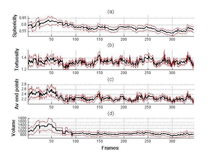
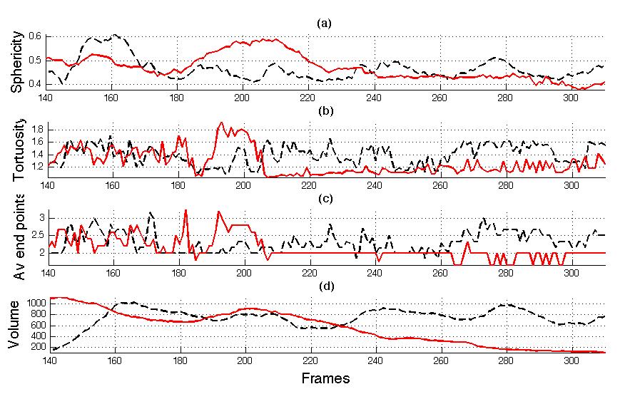
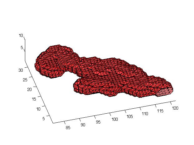
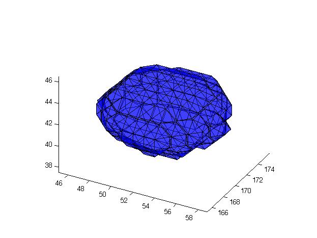
We have presented a paper at PRIB 2009 where a tracking and shape analysis algorithm for neutrophils of zebrafish is proposed. The neutrophils were fluorescently labelled with Green Fluorescent Protein and observed in a time-lapse three dimensional video through a confocal microscope.
The neutrophils were segmented from the background and
tracked with a keyhole model of movement. Morphological analysis was
performed by calculating the volume of the segmented objects together
with the measurements of sphericity, tortuosity and average number of
end points of the centre lines.
We speculate that these measurements are related to the activation of the neutrophils as part of the process of killing and digesting bacteria.
The algorithm is fully automatic and should provide a robust framework of analysis for posterior analysis of the neutrophils in zebrafish.