Measurement of vascular permeability
Vascular permeability of blood vessels in tumours is of
great
interest for the treatment of cancer for three main reasons. First, the
vascular wall represents a major barrier to the entry of high molecular
weight anti-cancer agents into tumour tissue; second, it controls the
tumour microenvironment thus affecting tumour progression and third,
changes in its barrier function may provide an early indicator of
vascular damage following treatment with anti-angiogenic or vascular
disrupting drugs. Despite the importance of tumour vascular
permeability, little attention has been paid to its analytic
measurement.

This algorithm was published in
Microcirculation.
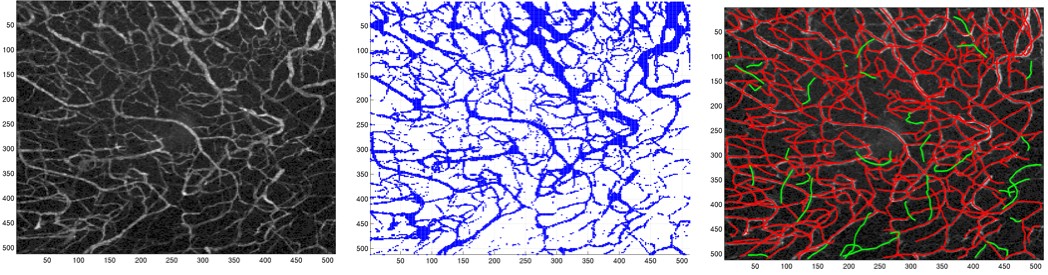
We have developed an image processing-based algorithm to measure the vascular permeability of microvessels to intravenously-administered 40 kDa fluorescein isothiocyanate (FITC) labelled dextran in tumours. The extravasation of the marker was imaged within P22 sarcomas growing in dorsal skin flap window chambers in BDIX rats using multiphoton fluorescence microscopy. Images of the tumour and its vasculature were obtained over a time-course of 1 hour in anaesthetized rats: 9 treated with the tumour vascular damaging agent, combretastatin A-4-phosphate (CA-4-P) and 6 controls. The algorithm segments the tissue and vessels in the volumetric data and measures voxel fluorescence intensity per class. The algorithm does not assume vessels as perfect cylinders nor involve the use of manually placed windows or regions of interest (ROI) as is commonly required. Instead, it considers all vessels in the data and therefore the average intensity measurement from a higher number of elements than those contained by a ROI becomes statistically more reliable. Patlak plots were used to determine average permeability-surface area product per unit volume (PS/V, x 10-4 sec-1) and permeability (P, x 10-7 cm/sec). The corresponding values for control and CA-4-P groups were 1.38+-0.71 and 2.64+-0.81 for PS/V (p=0.0049), and 1.31+-0.77 and 3.14+-1.58 for P (p=0.021). These results indicate: (i) statistical difference between controls and treated cases, (ii) higher vascular permeability for CA-4-P, and (iii) permeability values in the same range as those reported previously.