Measuring the Velocity of Fluorescently Labelled Red Blood Cells with a Keyhole Tracking Algorithm
We have proposed a tracking algorithm to measure the velocity
of
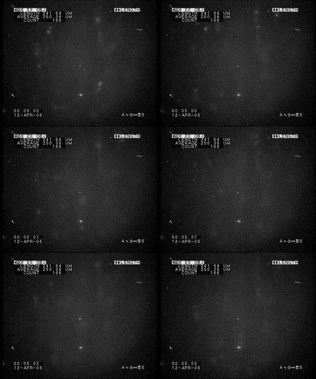
fluorescently labelled red blood cells (RBC) travelling through
microvessels of tumours, growing in dorsal skin flap window chambers,
implanted on mice.
This algorithm was published in the Journal of Microscopy.
My presentation of the algorithm at the MEDICON conference in Ljubljana, Slovenia was filmed and you can watch the video at Videolectures.net:
![]()
Measuring Red Blood Cell Velocity with a Keyhole Tracking Algorithm
Constantino Reyes-Aldasoro.

Pre-processing removed noise and
artefacts from the images and
then segmented cells from background.
The tracking algorithm is based
on a ‘keyhole’ model that
describes the probable movement of a segmented cell between contiguous
frames of a video sequence. When a history of cell movement exists,
past, present and a predicted landing position of the cells will define
two regions of probability that resemble the shape of a keyhole.
This
keyhole model was used to determine if cells in contiguous frames
should be linked to form tracks and also as a post-processing tool to
join split tracks and discard links that could have been formed due to
noise or uncertainty. When there was no history, a circular region
around the centroid of the parent cell was used as a region of
probability. Outliers were removed based on the distribution of the
average velocities of the tracks. Since the position and time of each
cell is recorded, a wealth of statistical measures can be obtained from
the tracks.
In one of the tumours there is a
complete
shutdown of the vasculature while in the other there is a clear
decrease of velocity at 30 minutes, with subsequent recovery by 6
hours. The tracking algorithm enabled the simultaneous measurement of
RBC velocity in multiple vessels within an intravital video sequence,
enabling analysis of heterogeneity of flow and response to treatment in
mouse models of cancer.


